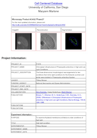
Microscopy product ID: 3432
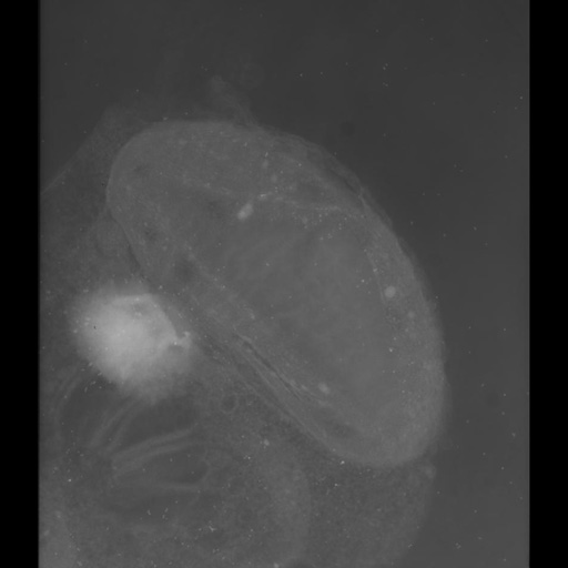
2D Image
| Description | Zero tilt image of a 0.5 um thick section of a blue green algae cell taken with intermediate voltage electron microscopy. Contrast is reversed so that electron dense structures appear bright. |
Embed
Embed URL
Embed Image
Full resolution data file
| File Size |
|
| File Format |
|
| Description | Tar file containing full resolution unaligned digitized images in suprim format (.f) along with the fiducial mark file used to align them (.fido) and the file containing a list of the angles used (.ang) and the origin file (.origin) |
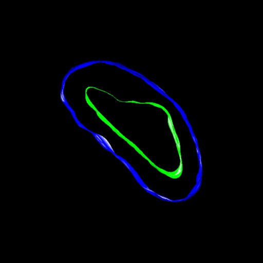
Segmentation
| Description | Manual segmentation of the chloroplast outer membrane and pyrenoid membrane using Xvoxtrace 2.7 followed by surfacing using Synu. Thylakoid membranes were difficult to resolve and so were not completely traced. |
Embed
Embed URL
Embed Image
Full resolution data file
| File Size |
|
| File Format |
|
| Description | Zipped archive containing the surfaced segmentations of the chloroplast outer membrane and the pyrenoid in Synu format along with the Viewdata file required for viewing with Synu. THe original trace file (.trace) containing the manual contours generated in Xvoxtrace is also included. |
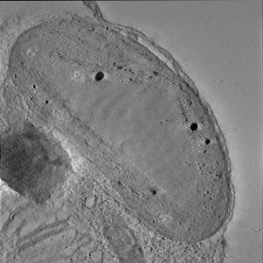
Reconstruction
| Description | Single computed slice of a tomographic volume of a chloroplast contained in a blue green algae cell grown under high light conditions. The pyrenoid is very prominent, although the thylakoid membranes are difficult to resolve. |
Embed
Embed URL
Embed Image
Full resolution data file
| File Size |
|
| File Format |
|
| Description | Tar file containing two volumes in Analyze 7.5 format: Obliques75.img/hdr and Phaeo27_91sub.img/hdr. Obliques75 was produced by reslicing Phaeo27_91sub along an oblique angle using the oblique section function of Analyze. |
Animation
Embed
Embed URL
Embed Video
| Description | Animation through the computed slices of a tomographic volume of a chloroplast contained in a blue green algae cell grown under high light conditions. The pyrenoid is very prominent, although the thylakoid membranes are difficult to resolve. |
Full metadata
- Collection
- Cite This Work
-
Moisan, Tiffany; Sosinsky, Gina; Buitenhuys, Casey; Ellisman, Mark H. (2017). Microscopy product ID: 3432. In Cell Centered Database. UC San Diego Library Digital Collections. https://doi.org/10.6075/J0P84BP1
- Date Issued
- 2017
- Research Team Head
- Researchers
- Technical Details
-
Product type: SINGLE TILT. Microscopy type: IVEM. Instrument: JEOL 4000EX IVEM
Strain: Karsten
- Funding
-
National Aeronautics and Space Administration
- Series
- Scientific Name
- Anatomy
- Topic
Formats
View formats within this collection
- Language
- No linguistic content; Not applicable
- Related Resources
- Moisan TA, Ellisman MH, Buitenhuys CW, Sosinsky GE (2006). Differences in chloroplast ultrastructure of Phaeocystis antarctica in low and high light. Marine Biology, 149(6):1281-1290. https://doi.org/10.1007/s00227-006-0321-5
- Microscopy product 3432 at the Cell Centered Database: https://doi.org/10.7295/W9CCDB3432
Primary associated publication
Other version
- License
-
Creative Commons Attribution 4.0 International Public License
- Rights Holder
- UC Regents
- Copyright
-
Under copyright (US)
Use: This work is available from the UC San Diego Library. This digital copy of the work is intended to support research, teaching, and private study.
Constraint(s) on Use: This work is protected by the U.S. Copyright Law (Title 17, U.S.C.). Use of this work beyond that allowed by "fair use" or any license applied to this work requires written permission of the copyright holder(s). Responsibility for obtaining permissions and any use and distribution of this work rests exclusively with the user and not the UC San Diego Library. Inquiries can be made to the UC San Diego Library program having custody of the work.
- Digital Object Made Available By
-
Research Data Curation Program, UC San Diego, La Jolla, 92093-0175 (https://lib.ucsd.edu/rdcp)
- Last Modified
2023-05-22