CIL:13875, Saccharomyces cerevisiae
CIL source metadata (JSON)
| File Size |
|
| File Format |
|
- Collection
- Cite This Work
-
Valerio-Santiago, Mauricio; Monje-Casas, Fernando (2021). CIL:13875, Saccharomyces cerevisiae. In Cell Image Library. UC San Diego Library Digital Collections. Dataset. https://doi.org/10.6075/J0FN14X5
- Description
-
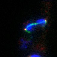
3HA-Bfa1 (red) localizes to both spindle pole bodies (SPBs) in anaphase in dyn1Δ cells with constitutive targeting of Tem1 to both SPBs. However, the constitutive presence of Tem1 on the SPBs impaired the spindle position checkpoint in the dyn1Δ cells. Anaphase was determined by spindle morphology (tubulin, green) and nuclear morphology (DAPI, blue). A DIC image is also shown (hidden). Image is Fig S3 in J Cell Biol. (2011) 192: 599-614.
- Date Issued
- 2021
- Researchers
- Methods
-
Cells (MATa tem1::KanMX ura3::eGFP-CNM67–TEM1::URA3 dyn1::URA3 bfa1::3HA-BFA1) grown in rich media at 14C for 24 hours were fixed for 15 min in 3.7% formaldehyde and 0.1 M potassium phosphate buffer, pH 6.4. Cells were then washed twice with 0.1 M potassium phosphate buffer, pH 6.4, and resuspended in 1.2 M sorbitol in 0.12 M K2HPO4/0.033 M citric acid, pH 5.9. Fixed cells were digested with 0.1 mg/ml zymolyase-100T (US Biological) and 1/10 volume of glusulase (PerkinElmer) at 30C for 15 min, washed once, and resuspended in 1.2 M sorbitol in 0.12 M K2HPO4/0.033 M citric acid, pH 5.9. Primary antibodies were anti-HA monoclonal antibody (HA.11; 1:500) and anti-tubulin (Abcam; 1:500). Secondary antibodies were: anti-mouse Cy3 (for HA) and anti-rat FITC (for tubulin). Cells were resuspended in DAPI (1 mg/ml). Imaging was performed at 25C using a Leica DM6000 microscope equipped with a 100x/1.40 NA oil immersion objective lens, A4, L5, and TX2 filters, and a digital CCD camera (DFC350, Leica). Pictures were processed with LAS AF (leica) and ImageJ software.
- Technical Details
-
Preparation: formaldehyde fixed tissue
Relation to intact cell: whole mounted tissue
Item type: recorded image
Imaging mode: widefield illumination; fluorescence microscopy; differential interference contrast microscopy
Parameter imaged: fluorescence emission; optical path length gradient
Source of contrast: distribution of a specific protein; compartmentalization of stain or label; boundaries between regions with different refractive index
Visualization methods: HA peptide tag; 4',6-diamidino-2-phenylindole (DAPI); primary antibody plus labeled secondary antibody; Cy3; Fluorescein (FITC)
Processing history: unprocessed raw data
Data qualification: Raw;spatialmeasurements;intensitiesquantitation - Series
- Scientific Name
- Anatomy
- Topics
-
- ATP binding
- Cell cycle
- Cell division
- Establishment of mitotic spindle orientation
- GTP binding
- GTPase activator activity
- GTPase activity
- Microtubule motor activity
- Microtubule-based movement
- Microtubule-based process
- Mitosis
- Mitotic cell cycle spindle orientation checkpoint
- Mitotic sister chromatid segregation
- Mitotic spindle elongation
- Nuclear migration along microtubule
- Nucleotide binding
- Protein binding
- Regulation of exit from mitosis
- Small GTPase mediated signal transduction
- Structural constituent of cytoskeleton
- W303
Formats
View formats within this collection
- Language
- No linguistic content; Not applicable
- Identifier
-
Samplenumber: 13875
- Related Resources
- Source Record in the Cell Image Library: https://doi.org/10.7295/W9CIL13875
- J Cell Biol. (2011) 192: 599-614. https://www.ncbi.nlm.nih.gov/pubmed/?term=21321099
- BioStudies (previously JCB DataViewer): https://www.ebi.ac.uk/biostudies/studies/
Source data
Reference
Other resource
- License
-
Creative Commons Attribution-NonCommercial-ShareAlike 4.0 International Public License
- Rights Holder
- UC Regents
- Copyright
-
Under copyright (US)
Use: This work is available from the UC San Diego Library. This digital copy of the work is intended to support research, teaching, and private study.
Constraint(s) on Use: This work is protected by the U.S. Copyright Law (Title 17, U.S.C.). Use of this work beyond that allowed by "fair use" or any license applied to this work requires written permission of the copyright holder(s). Responsibility for obtaining permissions and any use and distribution of this work rests exclusively with the user and not the UC San Diego Library. Inquiries can be made to the UC San Diego Library program having custody of the work.
- Digital Object Made Available By
-
Research Data Curation Program, UC San Diego, La Jolla, 92093-0175 (https://lib.ucsd.edu/rdcp)
- Last Modified
2022-08-12