Microscopy product ID: 8448
Reconstruction
| Description |
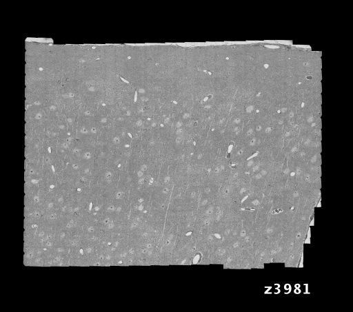
Serial electron micrograph series through layers 1-4 of the primary visual cortex of the adult mouse. Reconstruction encompasses 450 X 350 X 52 um volume of tissue and contains ~ 1500 cell bodies. This same area of tissue was functionally characterized for orientation and direction selectivity using two photon calcium imaging. |
Embed
Embed URL
Embed Image
Full resolution data file
| File Size |
|
| File Format |
|
| Description | Downsampled version (1/8 resolution) of a 10 Tb serial section reconstruction of mouse primary visual cortex available as a series of TIFF files. Each file is an assembled mosaic of a single thin section. A 2K X 2K full resolution core is also available for download under the "downsampled downloadable file" link. Note: The full 10 TB data set is available but may not be downloaded via the CCDB. If you are interested in accessing these data, please contact webmaster@ccdb.ucsd.edu Voxel dimensions given above for reconstruction represent values for the downsampled reconstruction. The original voxel dimensions are available under Image 2D or Microscopy Product tables. Authors note that the range of section thicknesses varied from 40 nm to 50 nm. An average value of 45 nm (0.045 um) was chosen here. |
Down sampled data file
| File Size |
|
| File Format |
|
| Description | Cropped version (2048 X 2048) of central core of serial electron micrograph series at full resolution (4 nm sampling) through layers 1-4 of the primary visual cortex of the adult mouse. Some of the 2k x 2k images are pure black. This is not a mistake; they are 'bookkeeping' sections, to maintain numbering alignment with the 1/8th resolution stack. If you look at the equivalent 1/8th section you will see that these are sections that have information outside the field of view of the 2k x 2k image. (These are 'coming in' sections. They occur after pickup of a series of sections, when the ultramicrotome is restarted and only grazes a portion of the sample block face for a few sections.) Note that the voxel dimensions of the cropped version are 0.004 um X 0.004 um X 0.045 um. |
Animation
Embed
Embed URL
Embed Video
| Description | Animation through the central core of serial electron micrograph series at full resolution (4 nm sampling) through layers 1-4 of the primary visual cortex of the adult mouse. Black images represent 'bookkeeping' sections inserted to maintain numbering alignment with the 1/8th resolution stack. The cropped full resolution volume used to create this animation is available for download through the "downsampled downloadable data file" link. |
Full metadata
- Collection
- Cite This Work
-
Reid, R. Clay; Bock, Davi; Lee, Wei-Chung Allen; Kerlin, Aaron M.; Andermann, Mark L.; Hood, Greg; Wetzel, Arthur W.; Yurgenson, Sergey; Soucy, Edward R.; Kim, Hyon Suk (2017). Microscopy product ID: 8448. In Cell Centered Database. UC San Diego Library Digital Collections. https://doi.org/10.6075/J0Z31ZFZ
- Date Issued
- 2017
- Research Team Head
- Researchers
- Technical Details
-
Male. Strain: Thy1-YFP-H
Product type: SERIAL SECTION. Microscopy type: TEM. Instrument: JEOL 1200EX
- Funding
-
National Eye Institute, National Institutes of Health
- Series
- Scientific Name
- Anatomy
- Topic
Formats
View formats within this collection
- Language
- No linguistic content; Not applicable
- Related Resources
- Bock DD, Lee WA, Kerlin AM, Andermann ML, Hood G, Wetzel AW, Yurgenson S, Soucy ER, Kim HS, Reid RC (2011). Network anatomy and in vivo physiology of visual cortical neurons. Nature, 471(7337):177-182. PubMed: https://www.ncbi.nlm.nih.gov/pmc/articles/PMC3095821/; Nature. https://doi.org/10.1038/nature09802
- Microscopy product 8448 at the Cell Centered Database: https://doi.org/10.7295/W9CCDB8448
Primary associated publication
Other version
- License
-
Creative Commons Attribution 4.0 International Public License
- Rights Holder
- UC Regents
- Copyright
-
Under copyright (US)
Use: This work is available from the UC San Diego Library. This digital copy of the work is intended to support research, teaching, and private study.
Constraint(s) on Use: This work is protected by the U.S. Copyright Law (Title 17, U.S.C.). Use of this work beyond that allowed by "fair use" or any license applied to this work requires written permission of the copyright holder(s). Responsibility for obtaining permissions and any use and distribution of this work rests exclusively with the user and not the UC San Diego Library. Inquiries can be made to the UC San Diego Library program having custody of the work.
- Digital Object Made Available By
-
Research Data Curation Program, UC San Diego, La Jolla, 92093-0175 (https://lib.ucsd.edu/rdcp)
- Last Modified
2023-05-22