CIL:10011, Didinium nasutum, eukaryotic cell, Eukaryotic Protist, Ciliated Protist
CIL source metadata (JSON)
| File Size |
|
| File Format |
|
- Collection
- Cite This Work
-
Allen, Richard (2021). CIL:10011, Didinium nasutum, eukaryotic cell, Eukaryotic Protist, Ciliated Protist. In Cell Image Library. UC San Diego Library Digital Collections. Dataset. https://doi.org/10.6075/J0SJ1JQH
- Description
-
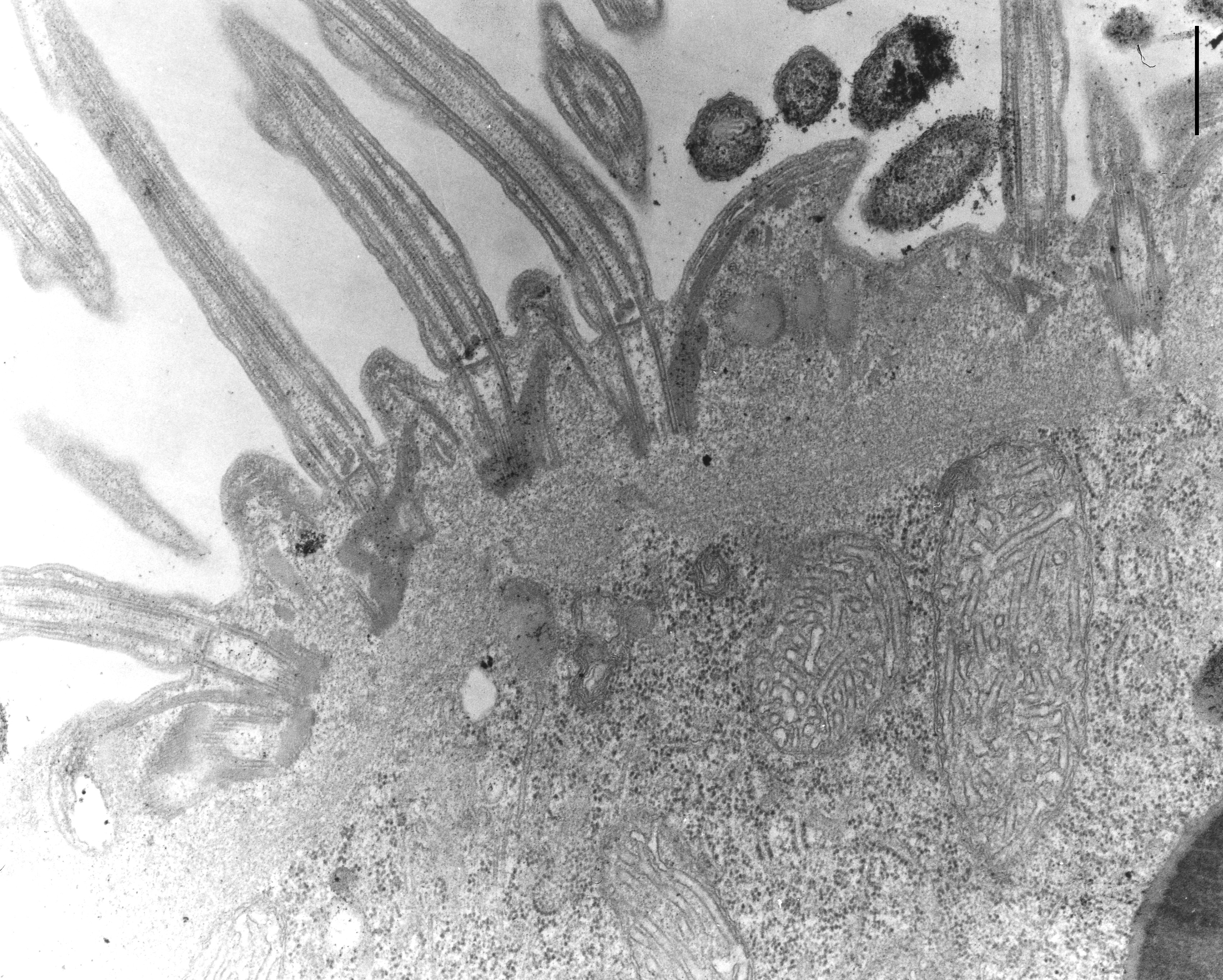
Didinium has two bands of ciliated basal bodies that encircle the cell, one adorally and one aborally. The bands are composed of groups of approximately 15 ciliated basal bodies. This section is a longitudinal section of the cilium and basal body and cross section of approximately seven different groups or pectinelles. The proximal end of basal bodies are ensheathed in electron opaque sleeves from which microtubular ribbons extend into the adjacent ridges on the two sides of each basal body. TEM taken on 5/20/69 by R. Allen with Philips 300 operating at 60kV. Neg. 14,800X.Bar = 0.5µm. The negative was printed to paper and the image was scanned to Photoshop. This digitized image is available for qualitative analysis. A raw, unprocessed, high resolution version of this image (CIL:4664) is in the library and available for quantitative analysis. Standard glutaraldehyde fixation followed by osmium tetroxide, dehydrated in alcohol and embedded in an epoxy resin. Microtome sections prepared at approximately 75nm thickness. Additional information available at (http://www5.pbrc.hawaii.edu/allen/).
- Date Issued
- 2021
- Researcher
- Technical Details
-
Preparation: glutaraldehyde fixed tissue; osmium tetroxide fixed tissue; tissue in epoxy resin embedment; microtome-sectioned tissue
Relation to intact cell: microtome-sectioned tissue
Item type: transmission electron microscopy (TEM); illumination by electrons
Imaging mode: detection of electrons; film
Parameter imaged: electron density
Source of contrast: stain with broad specificity
Visualization methods: stain with broad specificity; osmium tetroxide; uranyl salt; lead salt
Processing history: film; Print from negative scanned for Photoshop.
Data qualification: Processed;spatialmeasurements - Series
- Scientific Name
- Anatomy
- Topics
Formats
View formats within this collection
- Language
- No linguistic content; Not applicable
- Identifier
-
Samplenumber: 10011
- Related Resources
- Source Record in the Cell Image Library: https://doi.org/10.7295/W9CIL10011
- Paramecium and Other Ciliates: Richard Allen's Image Collection. Pacific Biosciences Research Center. University of Hawaii at Manoa: https://www5.pbrc.hawaii.edu/allen/
Source data
Other resource
- License
- Copyright
-
Creative Commons Public Domain Dedication
Use: The person(s) who associated a work with this deed has dedicated the work to the public domain by waiving all of their rights to the work worldwide under copyright law, including all related and neighboring rights, to the extent allowed by law.
Constraint(s) on Use: This work may be used without prior permission.
- Digital Object Made Available By
-
Research Data Curation Program, UC San Diego, La Jolla, 92093-0175 (https://lib.ucsd.edu/rdcp)
- Last Modified
2022-08-12