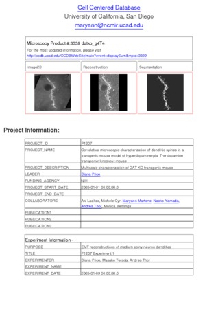
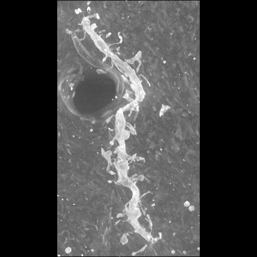
Microscopy product ID: 3339
2D Image
| Description | Electron micrograph of a selectively-stained spiny dendrite from a medium spiny neuron contained in a 4 um thick section from a dopamine transporter knock out mouse, imaged using ultra high voltage electron microscopy. |
Embed
Embed URL
Embed Image
Full resolution data file
| File Size |
|
| File Format |
|
| Description | Tar file containing IMOD files (datko_g4T4.com/.log/.st/.preali/.fid/.rawtlt) used for the alignment and the original tiff images (in the TIFF folder in the format datkoc_g4T4000.tif.gz) |
Animation
Embed
Embed URL
Embed Video
| Description | Animation of aligned electron microscopic tilt series of a selectively-stained spiny dendrite from a medium spiny neuron contained in a 4 um thick section from a dopamine transporter knock out mouse, imaged using ultra high voltage electron microscopy. Tilt series was obtained at 2 degree increments through +/- 70 degrees of tilt. |
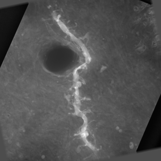
Segmentation
| Description | Segmentation of individual dendritic spines and shaft in Analyze .obj format. Spines were segmented using simple thresholding and morphological operations. |
Embed
Embed URL
Embed Image
Full resolution data file
| File Size |
|
| File Format |
|
| Description | a .tar file containing the .obj file generated by Analyze AVW (datko_g4T4.obj) along with the segmented volume in Analyze 7.5 format (datko_g4T4_mor.hdr/img). |
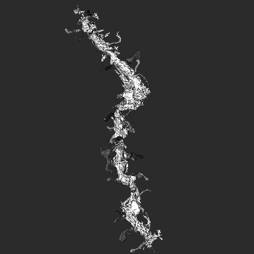
Reconstruction
| Description | Single computed slice through a tomographic reconstruction of a selectively-stained spiny dendrite from a medium spiny neuron contained in a 4 um thick section from the neostriatum of a dopamine transporter knock out mouse. |
Embed
Embed URL
Embed Image
Full resolution data file
| File Size |
|
| File Format |
|
| Description | Tar file containing the IMOD .rec format (datko_g4T4_full.rec) for the volume reconstruction and Amira .am/.hx format image stack of the labels used (datko_g4T4_full-labels.am, datko_g4T4.hx). |
Animation
Embed
Embed URL
Embed Video
| Description | A .qt movie of a maximum intensity projection from a spiny dendrite volume reconstruction. |
Full metadata
- Collection
- Cite This Work
-
Price, Diana; Laakso, Aki; Cyr, Michele; Martone, Maryann; Yamada, Naoko; Thor, Andrea; Berlanga, Monica L. (2017). Microscopy product ID: 3339. In Cell Centered Database. UC San Diego Library Digital Collections. https://doi.org/10.6075/J0R78F03
- Creation Date
- Microscopy product: 2003-12-22. Experiment: 2003-01-09. Project: 2003-01-01.
- Date Issued
- 2017
- Research Team Head
- Researchers
- Technical Details
-
Male. Strain: C57BL/129SvJ
Product type: single tilt tomography. Microscopy type: UHVEM. Instrument: Hitachi 3MeV UHVEM
- Funding
-
NIH
- Series
- Scientific Name
- Anatomy
- Topic
Formats
View formats within this collection
- Language
- No linguistic content; Not applicable
- Related Resource
- Microscopy product 3339 at the Cell Centered Database: https://doi.org/10.7295/W9CCDB3339
Other version
- License
-
Creative Commons Attribution 4.0 International Public License
- Rights Holder
- UC Regents
- Copyright
-
Under copyright (US)
Use: This work is available from the UC San Diego Library. This digital copy of the work is intended to support research, teaching, and private study.
Constraint(s) on Use: This work is protected by the U.S. Copyright Law (Title 17, U.S.C.). Use of this work beyond that allowed by "fair use" or any license applied to this work requires written permission of the copyright holder(s). Responsibility for obtaining permissions and any use and distribution of this work rests exclusively with the user and not the UC San Diego Library. Inquiries can be made to the UC San Diego Library program having custody of the work.
- Digital Object Made Available By
-
Research Data Curation Program, UC San Diego, La Jolla, 92093-0175 (https://lib.ucsd.edu/rdcp)
- Last Modified
2023-05-22