Microscopy product ID: 3652
2D Image
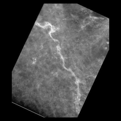
| Description | Electron micrograph of a spiny dendrite from medium spiny neurons in a dopamine transporter knock out mouse using diolistic labeling, imaged using ultra high voltage electron microscopy. |
Embed
Embed URL
Embed Image
Full resolution data file
| File Size |
|
| File Format |
|
| Description | Tar file containing IMOD files (tg6dkg14a.com/.log/.st/.preali/.fid/.rawtlt) used for the alignment and the original tiff images (in the TIFF folder in the format tg6dkg14a000.tif.gz) |
Animation
Embed
Embed URL
Embed Video
| Description | Animation of aligned electron microscopic tilt series of a spiny dendrite from medium spiny neurons in a dopamine transporter knock out mouse using diolistic labeling, imaged using ultra high voltage electron microscopy. Tilt series was obtained at 2 degree increments through +/-66 degrees of tilt. |
Full resolution data file
| File Size |
|
| File Format |
|
| Description | Tar file containing Xvoxtrace files (tg6dkg14a.trace and viewdata), Synu files (shaft.synu, Spine_01.synu, Spine_02.synu, etc.) and the Analyze volume (tg6dkg14a_res2.img/.hdr) for the segmentation. Also includes the image map file in tiff format (tg6dkg14a.imagemap.tiff). |
Reconstruction
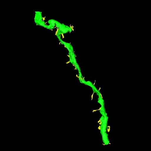
| Description | Electron micrograph of of spiny dendrites from medium spiny neurons in a dopamine transporter knock out mouse using diolistic labeling, imaged using ultra high voltage electron microscopy. |
Embed
Embed URL
Embed Image
Full resolution data file
| File Size |
|
| File Format |
|
| Description | Tar file containing the the IMOD .hdr/.img files for the of the electron tomograph (tg6dkg14a_an_sub.hdr/.img) |
Animation
Embed
Embed URL
Embed Video
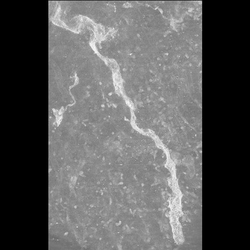
| Description | Animation of aligned electron microscopic tilt series of a spiny dendrite from medium spiny neurons in a dopamine transporter knock out mouse using diolistic labeling, imaged using ultra high voltage electron microscopy. Tilt series was obtained at 2 degree increments through +/- 66 degrees of tilt. Note: contrast has been reversed so that labeled dendrite appears bright and unstained tissue appears dark. |
Full metadata
- Collection
- Cite This Work
-
Price, Diana; Laakso, Aki; Cyr, Michele; Martone, Maryann; Yamada, Naoko; Thor, Andrea; Berlanga, Monica L. (2017). Microscopy product ID: 3652. In Cell Centered Database. UC San Diego Library Digital Collections. https://doi.org/10.6075/J0P55N95
- Creation Date
- Project: 2003-01-01.
- Date Issued
- 2017
- Research Team Head
- Researchers
- Technical Details
-
Product type: SINGLE TILT. Microscopy type: UHVEM. Instrument: Hitachi 3MeV UHVEM
Strain: B6;129-/Slc6a3 tm2Mca
- Funding
-
NIH
- Series
- Scientific Name
- Anatomy
- Topic
Formats
View formats within this collection
- Language
- No linguistic content; Not applicable
- Related Resource
- Microscopy product 3652 at the Cell Centered Database: https://doi.org/10.7295/W9CCDB3652
Other version
- License
-
Creative Commons Attribution 4.0 International Public License
- Rights Holder
- UC Regents
- Copyright
-
Under copyright (US)
Use: This work is available from the UC San Diego Library. This digital copy of the work is intended to support research, teaching, and private study.
Constraint(s) on Use: This work is protected by the U.S. Copyright Law (Title 17, U.S.C.). Use of this work beyond that allowed by "fair use" or any license applied to this work requires written permission of the copyright holder(s). Responsibility for obtaining permissions and any use and distribution of this work rests exclusively with the user and not the UC San Diego Library. Inquiries can be made to the UC San Diego Library program having custody of the work.
- Digital Object Made Available By
-
Research Data Curation Program, UC San Diego, La Jolla, 92093-0175 (https://lib.ucsd.edu/rdcp)
- Last Modified
2023-05-22