Microscopy product ID: 6348
2D Image
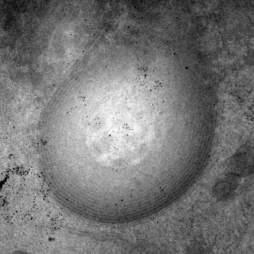
| Description | Single tilt image taken at zero degrees tilt of a lamellar structure observed in HeLa cells transfected with mutant connexin 50, imaged using intermediate voltage electron microscopy, and labeled using the tetracysteine-ReHAsH system and fluorescence photoooxidation. The concentric double membrane layers of the aggregate appear to form "onion skin" sheets that do not form a closed structure along the long axis. The differential staining between the outside and inside is most likely due to impeded diffusion of DAB into the interior of these accumulations. Dark particles represent 20 nm gold particles applied to the surface of the section to serve as fiducial marks for subsequent alignment. |
Embed
Embed URL
Embed Image
Full resolution data file
| File Size |
|
| File Format |
|
| Description | Zipped archive containing the tilt images in IMOD format along with all supporting IMOD files for alignment (beyers2a_*). |
Animation
Embed
Embed URL
Embed Video
| Description | Animation through the aligned sections of a tilt series through a lamellar structure observed in HeLa cells transfected with mutant connexin 50, imaged using intermediate voltage electron microscopy, and labeled using the tetracysteine-ReHAsH system and fluorescence photoooxidation. The concentric double membrane layers of the aggregate appear to form "onion skin" sheets that do not form a closed structure along the long axis. The differential staining between the outside and inside is most likely due to impeded diffusion of DAB into the interior of these accumulations. Dark particles represent 20 nm gold particles applied to the surface of the section to serve as fiducial marks for subsequent alignment. |
Segmentation
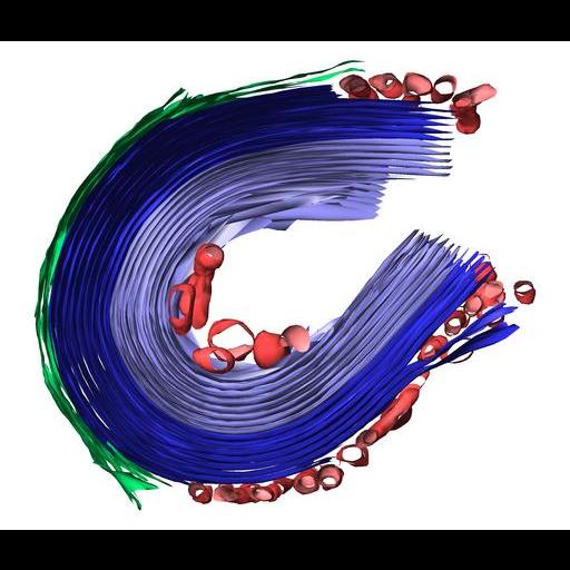
| Description | Manual segmentation of CX50 labeled lamellae, rough endoplasmic reticulum and vesicles using IMOD. Segmentations were surface rendered using Amira. |
Embed
Embed URL
Embed Image
Full resolution data file
| File Size |
|
| File Format |
|
| Description | Gzipped, tar file which contains: Volume used to produce model file(s), IMOD model file containing three objects (each with many contours), model files of each contour in IMOD and VRML format, Amira network used to create surfaced models and movies. |
Reconstruction
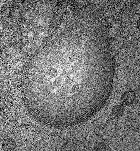
| Description | Single computed slice through a double tilt electron tomogram of a lamellar structure observed in HeLa cells transfected with mutant connexin 50, imaged using intermediate voltage electron microscopy, and labeled using the tetracysteine-ReHAsH system and fluorescence photoooxidation. The concentric double membrane layers of the aggregate appear to form "onion skin" sheets that do not form a closed structure along the long axis. The differential staining between the outside and inside is most likely due to impeded diffusion of DAB into the interior of these accumulations. |
Embed
Embed URL
Embed Image
Full resolution data file
| File Size |
|
| File Format |
|
| Description | Double tilt tomographic volume file in IMOD (MRC) format. |
Animation
Embed
Embed URL
Embed Video
| Description | Animation through the computed slices of a double tilt electron tomogram through a Single computed slice through a double tilt electron tomogram of a lamellar structure observed in HeLa cells transfected with mutant connexin 50, imaged using intermediate voltage electron microscopy, and labeled using the tetracysteine-ReHAsH system and fluorescence photoooxidation. The concentric double membrane layers of the aggregate appear to form "onion skin" sheets that do not form a closed structure along the long axis. The differential staining between the outside and inside is most likely due to impeded diffusion of DAB into the interior of these accumulations. |
Full metadata
- Collection
- Cite This Work
-
Beyer, Eric; Sosinsky, Gina; Crum, John; Berthoud, Viviana; Lichtensetin, Alexandra; Gaietta, Guido (2017). Microscopy product ID: 6348. In Cell Centered Database. UC San Diego Library Digital Collections. https://doi.org/10.6075/J0Q2401M
- Date Issued
- 2017
- Research Team Head
- Researchers
- Technical Details
-
Female
Product type: DOUBLE TILT. Microscopy type: IVEM. Instrument: JEOL 4000EX IVEM
- Funding
-
National Eye Institute, National Institutes of Health
- Series
- Scientific Name
- Anatomy
- Topic
Formats
View formats within this collection
- Language
- No linguistic content; Not applicable
- Related Resources
- Lichtenstein A, Gaietta GM, Deerinck TJ, Crum J, Sosinsky GE, Beyer EC, Berthoud VM (2009). The cytoplasmic accumulations of the cataract-associated mutant, Connexin50P88S, are long-lived and form in the endoplasmic reticulum. Experimental Eye Research, 88(3):600-609. PubMed: https://www.ncbi.nlm.nih.gov/pmc/articles/PMC2695785/; Experimental Eye Research. https://doi.org/10.1016/j.exer.2008.11.024
- Microscopy product 6348 at the Cell Centered Database: https://doi.org/10.7295/W9CCDB6348
Primary associated publication
Other version
- License
-
Creative Commons Attribution 4.0 International Public License
- Rights Holder
- UC Regents
- Copyright
-
Under copyright (US)
Use: This work is available from the UC San Diego Library. This digital copy of the work is intended to support research, teaching, and private study.
Constraint(s) on Use: This work is protected by the U.S. Copyright Law (Title 17, U.S.C.). Use of this work beyond that allowed by "fair use" or any license applied to this work requires written permission of the copyright holder(s). Responsibility for obtaining permissions and any use and distribution of this work rests exclusively with the user and not the UC San Diego Library. Inquiries can be made to the UC San Diego Library program having custody of the work.
- Digital Object Made Available By
-
Research Data Curation Program, UC San Diego, La Jolla, 92093-0175 (https://lib.ucsd.edu/rdcp)
- Last Modified
2023-05-22