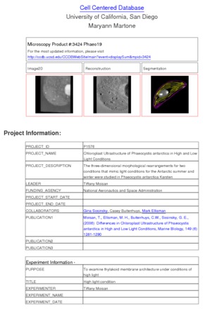
Microscopy product ID: 3424
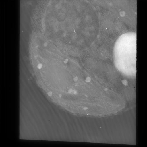
2D Image
| Description | Zero degree tilt image of a 0.25 um thick section of blue green algae grown under high light conditions, imaged with intermediate voltage electron microscopy. Contrast is reversed so that electron dense structures appear bright. |
Embed
Embed URL
Embed Image
Full resolution data file
| File Size |
|
| File Format |
|
| Description | Zip file containing the compressed full size digitized unaligned images (*.f.gz) in suprim format, along with the fiducial mark file used to align them (*.fido), the origin file (*.origin) and a list of the angles used for reconstruction (*.ang). |
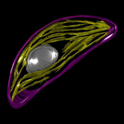
Segmentation
| Description | Manual segmentation of thylakoid membranes and other cellular structures using Xvoxtrace 2.1, followed by surfacing using the nuages algorithm and Synu. |
Embed
Embed URL
Embed Image
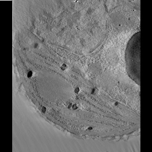
Reconstruction
| Description | Computed slice through tomographic reconstruction of blue green algae grown under high light conditions. |
Embed
Embed URL
Embed Image
Full resolution data file
| File Size |
|
| File Format |
|
| Description | Zip file containing volume file in Analyze 7.5 format (both .img and .hdr) files, used for segmentation. |
Animation
Embed
Embed URL
Embed Video
| Description | Animation through the computed slices of a tomographic reconstruction of a blue green algae grown under high light conditions. |
Full metadata
- Collection
- Cite This Work
-
Moisan, Tiffany; Sosinsky, Gina; Buitenhuys, Casey; Ellisman, Mark H. (2017). Microscopy product ID: 3424. In Cell Centered Database. UC San Diego Library Digital Collections. https://doi.org/10.6075/J06973C7
- Date Issued
- 2017
- Research Team Head
- Researchers
- Technical Details
-
Product type: SINGLE TILT. Microscopy type: IVEM. Instrument: JEOL 4000EX IVEM
Strain: Karsten
- Funding
-
National Aeronautics and Space Administration
- Series
- Scientific Name
- Anatomy
- Topic
Formats
View formats within this collection
- Language
- No linguistic content; Not applicable
- Related Resources
- Moisan TA, Ellisman MH, Buitenhuys CW, Sosinsky GE (2006). Differences in chloroplast ultrastructure of Phaeocystis antarctica in low and high light. Marine Biology, 149(6):1281-1290. https://doi.org/10.1007/s00227-006-0321-5
- Microscopy product 3424 at the Cell Centered Database: https://doi.org/10.7295/W9CCDB3424
Primary associated publication
Other version
- License
-
Creative Commons Attribution 4.0 International Public License
- Rights Holder
- UC Regents
- Copyright
-
Under copyright (US)
Use: This work is available from the UC San Diego Library. This digital copy of the work is intended to support research, teaching, and private study.
Constraint(s) on Use: This work is protected by the U.S. Copyright Law (Title 17, U.S.C.). Use of this work beyond that allowed by "fair use" or any license applied to this work requires written permission of the copyright holder(s). Responsibility for obtaining permissions and any use and distribution of this work rests exclusively with the user and not the UC San Diego Library. Inquiries can be made to the UC San Diego Library program having custody of the work.
- Digital Object Made Available By
-
Research Data Curation Program, UC San Diego, La Jolla, 92093-0175 (https://lib.ucsd.edu/rdcp)
- Last Modified
2023-05-22