Microscopy product ID: 3611
Segmentation
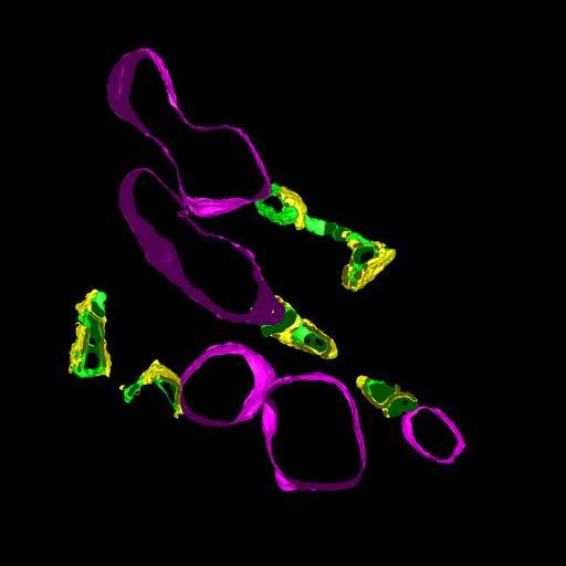
| Description | Manual segmentation of T tubule and SR membranes and mitochondria using IMOD 3.9.3 |
Embed
Embed URL
Embed Image
Full resolution data file
| File Size |
|
| File Format |
|
| Description | file contains IMOD generated tomographic model file (WT5aNC1_CCDB.mod) and information file that contains measurements from the segmentations generated by IMOD (WT5aNC1_CCDB.txt) |
Reconstruction
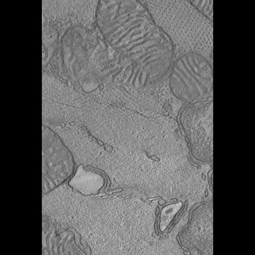
| Description | Portion of a single computed slice through a tomographic reconstruction of the myocardium of the left ventricle of a normal mouse. Note that the full reconstruction covers a larger area than is displayed here. |
Embed
Embed URL
Embed Image
Full resolution data file
| File Size |
|
| File Format |
|
| Description | Full resolution reconstruction by TxBR (quadratic: WT5aNC1_quad.rec) (~50 Gb). Note that a binned down reconstruction was used for segmentation that is available for download through the Downsampled data file download link. |
Down sampled data file
| File Size |
|
| File Format |
|
| Description | 2X binned down version of the reconstruction file in IMOD format. This version was used to generate the segmentation files. Note: to open this file using Jinx, append a .rec to the file name. |
Animation
Embed
Embed URL
Embed Video
| Description | Animation through the computed slices of a tomographic reconstruction from the left ventricle of a normal mouse heart. The electron dense particles on the surface of the volume are colloidal particles applied to the surface of the imaged section to serve as fiducial markers for alignment of the tilt series. |
Full metadata
- Collection
- Cite This Work
-
Hoshijima, Masahiko; Hayashi, Takeharu; Thor, Andrea; Terada, Masako; Martone, Maryann; Ellisman, Mark H. (2017). Microscopy product ID: 3611. In Cell Centered Database. UC San Diego Library Digital Collections. https://doi.org/10.6075/J0ZK5GFH
- Creation Date
- Project: 2004-04-01.
- Date Issued
- 2017
- Research Team Heads
- Researchers
- Technical Details
-
Male. Strain: S129
Product type: SINGLE TILT. Microscopy type: IVEM. Instrument: JEOL 4000 #1
- Series
- Scientific Name
- Anatomy
- Topic
Formats
View formats within this collection
- Language
- No linguistic content; Not applicable
- Related Resources
- Hayashi T, Martone ME, Yu Z, Thor A, Doi M, Holst MJ, Ellisman MH, Hoshijima M (2009). Three-dimensional electron microscopy reveals new details of membrane systems for calcium signaling in the heart. J. Cell Science, 122:1005-1013. PubMed: https://www.ncbi.nlm.nih.gov/pmc/articles/PMC2720931/; Journal of Cell Science. https://doi.org/10.1242/jcs.028175
- Microscopy product 3611 at the Cell Centered Database: https://doi.org/10.7295/W9CCDB3611
Primary associated publication
Other version
- License
-
Creative Commons Attribution 4.0 International Public License
- Rights Holder
- UC Regents
- Copyright
-
Under copyright (US)
Use: This work is available from the UC San Diego Library. This digital copy of the work is intended to support research, teaching, and private study.
Constraint(s) on Use: This work is protected by the U.S. Copyright Law (Title 17, U.S.C.). Use of this work beyond that allowed by "fair use" or any license applied to this work requires written permission of the copyright holder(s). Responsibility for obtaining permissions and any use and distribution of this work rests exclusively with the user and not the UC San Diego Library. Inquiries can be made to the UC San Diego Library program having custody of the work.
- Digital Object Made Available By
-
Research Data Curation Program, UC San Diego, La Jolla, 92093-0175 (https://lib.ucsd.edu/rdcp)
- Last Modified
2023-05-22