Microscopy product ID: 6739
2D Image
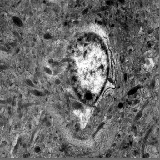
| Description | Electron micrograph of the soma of a protoplasmic astrocyte in a 0.5 um thick section from the hippocampus of a 1 month old male mouse, imaged with intermediate voltage electron microscopy at zero degrees tilt. This section is the 19th in a series of 26 serial semi-thick sections through the astrocyte soma used for a serial tomographic reconstruction (MP7503). |
Embed
Embed URL
Embed Image
Full resolution data file
| File Size |
|
| File Format |
|
| Description | File contains all IMOD and TxBR files (st, fid, preali, *com, *log, *mat, etc...) |
Animation
Embed
Embed URL
Embed Video
| Description | Animated tilt series of the soma of a protoplasmic astrocyte in a 0.5 um thick section from the hippocampus of a 1 month old male mouse, imaged with intermediate voltage electron microscopy. This section is the 19th in a series of 26 serial semi-thick sections through the astrocyte soma used for a serial tomographic reconstruction (MP7503). |
Reconstruction
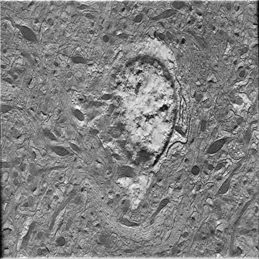
| Description | Single computed slice through a tomographic reconstruction of a protoplasmic astrocyte in a 0.5 um thick section from the hippocampus of a 1 month old male mouse, imaged with intermediate voltage electron microscopy. This reconstruction is the 19th in a series of 26 serial reconstructions through the cell soma. The complete reconstruction can be viewed under MP7503. |
Embed
Embed URL
Embed Image
Full resolution data file
| File Size |
|
| File Format |
|
| Description | File contains TxBR generated tomographic reconstructions in IMOD MRC format |
Down sampled data file
| File Size |
|
| File Format |
|
| Description | File contains binned down TxBR generated tomographic reconstructions binned by a factor of 8. Note that stated resolution is for full resolution volume and not for the downsampled volume. |
Animation
Embed
Embed URL
Embed Video
| Description | Animation through the computed slices of a tomographic reconstruction of a protoplasmic astrocyte in a 0.5 um thick section from the hippocampus of a 1 month old male mouse, imaged with intermediate voltage electron microscopy. This reconstruction is the 19th in a series of 26 serial reconstructions through the cell soma. The complete reconstruction can be viewed under MP7503. |
Full metadata
- Collection
- Cite This Work
-
Ellisman, Mark H.; Larson, Stephen; Maynard, Sarah; Bushong, Eric A.; Martone, Maryann (2017). Microscopy product ID: 6739. In Cell Centered Database. UC San Diego Library Digital Collections. https://doi.org/10.6075/J0ZC82N9
- Creation Date
- Experiment: 2009-10-01. Project: 2009-08-05.
- Date Issued
- 2017
- Research Team Heads
- Researchers
- Technical Details
-
Male. Strain: C57BL/6NHsd
Product type: SINGLE TILT. Microscopy type: IVEM. Instrument: JEOL 4000 #2
- Funding
-
Ted Waitt Family Foundation
- Series
- Scientific Name
- Anatomy
- Topic
Formats
View formats within this collection
- Language
- No linguistic content; Not applicable
- Related Resource
- Microscopy product 6739 at the Cell Centered Database: https://doi.org/10.7295/W9CCDB6739
Other version
- License
-
Creative Commons Attribution 4.0 International Public License
- Rights Holder
- UC Regents
- Copyright
-
Under copyright (US)
Use: This work is available from the UC San Diego Library. This digital copy of the work is intended to support research, teaching, and private study.
Constraint(s) on Use: This work is protected by the U.S. Copyright Law (Title 17, U.S.C.). Use of this work beyond that allowed by "fair use" or any license applied to this work requires written permission of the copyright holder(s). Responsibility for obtaining permissions and any use and distribution of this work rests exclusively with the user and not the UC San Diego Library. Inquiries can be made to the UC San Diego Library program having custody of the work.
- Digital Object Made Available By
-
Research Data Curation Program, UC San Diego, La Jolla, 92093-0175 (https://lib.ucsd.edu/rdcp)
- Last Modified
2023-05-22