Microscopy product ID: 3632
2D Image
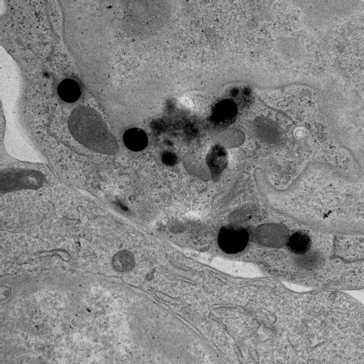
| Description | Projection of the untilted specimen: immunological synapse between a human cytotoxix T lymphocyte (top) and a target cell (bottom). |
Embed
Embed URL
Embed Image
Full resolution data file
| File Size |
|
| File Format |
|
| Description | The dual tilt axis series are stored as two MRC files (int, 16 bit): 1. CTL_ImmunSynapse_series_a.mrc, 2. CTL_ImmunSynapse_series_b.mrc The gziped tar file was created on a LINUX machine: tar -czf CTL_ImmunSynapse_img.tar.gz *mrc |
Segmentation
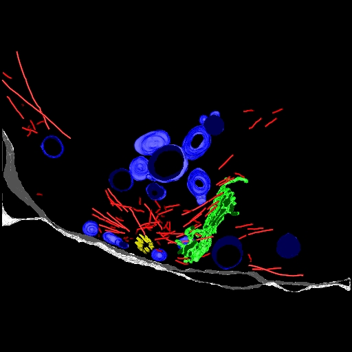
| Description | Segmentation of centriole, microtubules, Golgi apparatus and lytic granules within the CTL and of the synaptic cleft. |
Embed
Embed URL
Embed Image
Full resolution data file
| File Size |
|
| File Format |
|
| Description | Segmentation has been done with IMOD and AMIRA. The following objects have been segmented: 1. centriole (in the CTL), 2. microtubules (in the CTL), 3. Golgi apparatus (in the CTL), 4. lytic granules (in the CTL), 5. synaptic cleft (between CTL and target cell). The final model is an IMOD model file: CTL_ImmunSynapse_IMOD.mod.gz (it is gziped on a LIMUX mashine: gzip CTL_ImmunSynapse_IMOD.mod). Masks representing the various objects were generated in MRC format (int, 8 bit): 1. CTL_ImmunSynapse_centriole.mrc.gz, 2. CTL_ImmunSynapse_microtubules.mrc.gz, 3. CTL_ImmunSynapse_Golgi.mrc.gz, 4. CTL_ImmunSynapse_granules.mrc.gz, 5. CTL_ImmunSynapse_synapse.mrc.gz (files are gziped on a LIMUX mashine: gzip CTL_ImmunSynapse_*.mrc). The tar file was created on a LINUX machine: tar -cf CTL_ImmunSynapse_seg.tar CTL_ImmunSynapse_*.gz |
Reconstruction
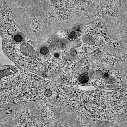
| Description | Tomogram of the immunological synapse between a cytotoxic T lymphocyte (CTL, top) and a target cell (bottom). The microtubule organization centre (MTOC) is polarized to the cell-cell contact site. The electron-dense lytic granules are transported along the microtubules to the synaptic cleft. Here they probably fuse with the membrane and excrete the cytotoxic proteins that trigger the death of the target cell. |
Embed
Embed URL
Embed Image
Full resolution data file
| File Size |
|
| File Format |
|
| Description | The tomographic reconstruction is stored as an MRC file (int, 8 bit): CTL_ImmunSynapse_rec.mrc.gz The tomogram has been Gauss-filtered with AMIRA (filter paramerters: 3D, Sigma x=y=z=1, Kernel x=y=z=3) as an MRC file (int, 8 bit): CTL_ImmunSynapse_filter_rec.mrc Segmentation was done using the filtered tomogram. The tar file was created on a LINUX machine: tar -cf CTL_ImmunSynapse_vol.tar *mrc |
Animation
Embed
Embed URL
Embed Video
| Description | Movie showing the tomogram of the immunological synapse between a cytotoxic T lymphocyte (CTL, top) and a target cell (bottom). The microtubule organization centre (MTOC) is polarized to the cell-cell contact site. The electron-dense lytic granules are transported along the microtubules to the synaptic cleft. Here they probably fuse with the membrane and excrete the cytotoxic proteins that trigger the death of the target cell. |
Full metadata
- Collection
- Cite This Work
-
Fuller, Stephen; Majorovits, Endre; Griffiths, Gillian; Stinchcombe, Jane; Bossi, Giovanna (2017). Microscopy product ID: 3632. In Cell Centered Database. UC San Diego Library Digital Collections. https://doi.org/10.6075/J09W0F85
- Creation Date
- Microscopy product: 2006-11-08. Experiment: 2005-11-08. Project: 2004-02-02.
- Date Issued
- 2017
- Research Team Heads
- Researchers
- Technical Details
-
Product type: DOUBLE TILT. Microscopy type: HVEM. Instrument: FEI Tecnai F30
- Funding
-
Wellcome Trust
- Series
- Scientific Name
- Anatomy
- Topic
Formats
View formats within this collection
- Language
- No linguistic content; Not applicable
- Related Resources
- Martone ME, Jones YZ, Young SJ, Ellisman MH, Zivin JA, Hu BR (1999). Modification of postsynaptic densities following transient cerebral ischemia: a quantitative and three dimensional ultrastructural study. J. Neurosci, 19(6):1988-1997. PubMed: https://www.ncbi.nlm.nih.gov/pubmed/10066252; Journal of Neuroscience. https://doi.org/10.1523/JNEUROSCI.19-06-01988.1999
- Microscopy product 3632 at the Cell Centered Database: https://doi.org/10.7295/W9CCDB3632
Primary associated publication
Other version
- License
-
Creative Commons Attribution 4.0 International Public License
- Rights Holder
- UC Regents
- Copyright
-
Under copyright (US)
Use: This work is available from the UC San Diego Library. This digital copy of the work is intended to support research, teaching, and private study.
Constraint(s) on Use: This work is protected by the U.S. Copyright Law (Title 17, U.S.C.). Use of this work beyond that allowed by "fair use" or any license applied to this work requires written permission of the copyright holder(s). Responsibility for obtaining permissions and any use and distribution of this work rests exclusively with the user and not the UC San Diego Library. Inquiries can be made to the UC San Diego Library program having custody of the work.
- Digital Object Made Available By
-
Research Data Curation Program, UC San Diego, La Jolla, 92093-0175 (https://lib.ucsd.edu/rdcp)
- Last Modified
2023-05-22