Microscopy product ID: 8244
2D Image
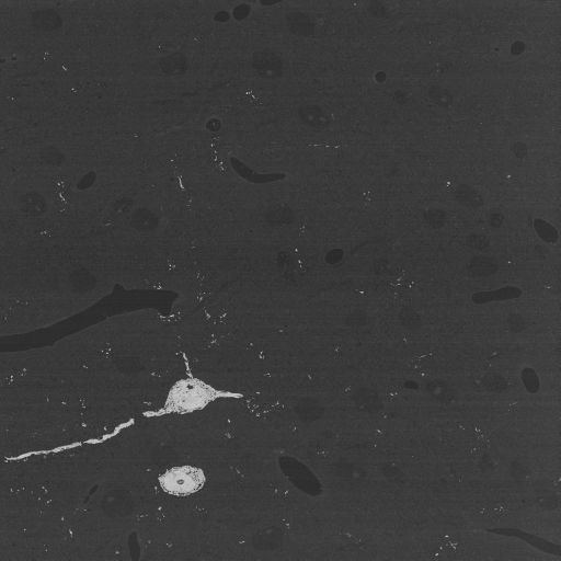
| Description | Single slice through Golgi-impregnated neurons from the mouse neocortex imaged using serial block face SEM. Contrast is reversed so that electron dense regions appear bright. |
Embed
Embed URL
Embed Image
Full resolution data file
| File Size |
|
| File Format |
|
| Description | Tar file containing raw data files in dm3 format |
Animation
Embed
Embed URL
Embed Video
| Description | Slice by slice animation of Golgi-impregnated neurons from the mouse neocortex imaged using serial block face SEM. Contrast is reversed so that electron dense regions appear bright. |
Segmentation
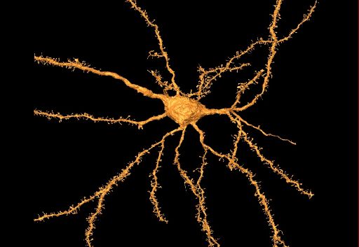
| Description | Semi automated segmentation of Golgi stained pyramidal neurons in the cerebral cortex using Amira |
Embed
Embed URL
Embed Image
Reconstruction
| Description | Golgi impregnated pyramidal neurons in the neocortex of an adult mouse. Contrast is reversed so that Golgi stain appears bright against the unstained background. A maximum intensity projection of the dataset is available in the thumbnail and by clicking the JPEG tab. |
Embed
Embed URL
Embed Image
Full resolution data file
| File Size |
|
| File Format |
|
| Description | Full resolution file in IMOD format. The raw series had 1560 planes total, but there is no information available as to why some planes were left off the reconstruction. |
Down sampled data file
| File Size |
|
| File Format |
|
| Description | Volume binned by a factor of 2 in the x, y and z planes: 2048 X 2048 X 632 slices |
Animation
Embed
Embed URL
Embed Video
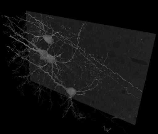
| Description | Animation showing the segmentation of individual neurons from a reconstruction of Golgi-impregnated mouse neocortex using serial block face SEM. Segmentation files are available for download under the segmentation section. |
Full metadata
- Collection
- Cite This Work
-
Ellisman, Mark H.; Larson, Stephen; Maynard, Sarah; Bushong, Eric A.; Martone, Maryann (2017). Microscopy product ID: 8244. In Cell Centered Database. UC San Diego Library Digital Collections. https://doi.org/10.6075/J02V2FXX
- Creation Date
- Microscopy product: 2009-09-03. Experiment: 2009-10-01. Project: 2009-08-05.
- Date Issued
- 2017
- Research Team Heads
- Researchers
- Technical Details
-
Male. Strain: C57BL/6
Product type: SERIAL SECTION. Microscopy type: Serial Block Face SEM. Instrument: FEI Quanta 200 FEG SEM with Gatan 3 View
- Funding
-
Ted Waitt Family Foundation
- Series
- Scientific Name
- Anatomy
- Topic
Formats
View formats within this collection
- Language
- No linguistic content; Not applicable
- Related Resource
- Microscopy product 8244 at the Cell Centered Database: https://doi.org/10.7295/W9CCDB8244
Other version
- License
-
Creative Commons Attribution 4.0 International Public License
- Rights Holder
- UC Regents
- Copyright
-
Under copyright (US)
Use: This work is available from the UC San Diego Library. This digital copy of the work is intended to support research, teaching, and private study.
Constraint(s) on Use: This work is protected by the U.S. Copyright Law (Title 17, U.S.C.). Use of this work beyond that allowed by "fair use" or any license applied to this work requires written permission of the copyright holder(s). Responsibility for obtaining permissions and any use and distribution of this work rests exclusively with the user and not the UC San Diego Library. Inquiries can be made to the UC San Diego Library program having custody of the work.
- Digital Object Made Available By
-
Research Data Curation Program, UC San Diego, La Jolla, 92093-0175 (https://lib.ucsd.edu/rdcp)
- Last Modified
2023-05-22