CIL:32146, Drosophila melanogaster, epithelial cell
CIL source metadata (JSON)
| File Size |
|
| File Format |
|
- Collection
- Cite This Work
-
Carpenter, Anne (2021). CIL:32146, Drosophila melanogaster, epithelial cell. In Cell Image Library. UC San Diego Library Digital Collections. Dataset. https://doi.org/10.6075/J02V2F35
- Description
-
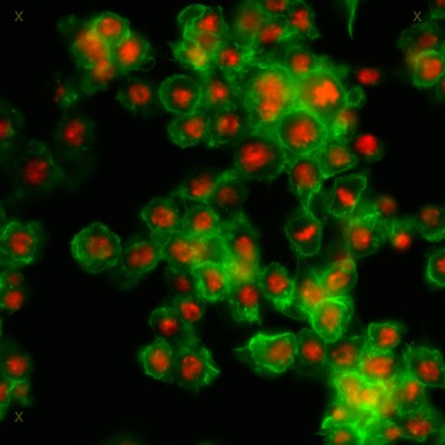
Drosophila melanogaster Kc167 cells were stained for DNA (to label nuclei, red) and actin (a cytoskeletal protein, to show the cell body, green). Each image is a dual channel fluorescent image followed by a line tracing of the nuclei and a line tracing of the cell boundary. These images are provided as a tool for testing algorithims to find nuclei and the boundaries between adjacent cells.
Chris Gang outlined the nuclei and cells.
Please use the following recommended citation when using this data. "We used the Drosophila Kc167 2 image set (Jones et al., Proc. ICCV Workshop on Computer Vision for Biomedical Image Applications, 2005), available from the Broad Bioimage Benchmark Collection (www.broad.mit.edu/bbbc)." - Date Issued
- 2021
- Researcher
- Methods
-
Images were acquired using a motorized Zeiss Axioplan 2 and a Axiocam MRm camera, and are provided courtesy of the laboratory of David Sabatini at the Whitehead Institute for Biomedical Research. Each image is roughly 512 x 512 pixels, with cells roughly 25 pixels in diameter, and 80 cells per image on average.
Algorithms for finding boundaries between adjacent cells may use the union of the manually outlined cells as input. They may also use the manually outlined nuclei as seed regions. This ensures that the algorithms are compared according to their ability to find boundaries betwen adjacent cells, and not the orthogonal problems of segmenting nuclei and distinguishing foreground (cells) from background.
To compare an algorithm's results to the manual outlines, define the relevant boundary pixels as the pixels that are on the boundary found by the algorith and that are not adjacent to any background pixels. For each relevant pixel, compute the Euclidean distance to the corresponding pixel on the manually found outline. Report the percentage of relevant pixels that are within two pixels of the corresponding pixel on the manually found outline. - Technical Details
-
Relation to intact cell: dispersed cells in vitro
Item type: recorded image; hand drawn contour
Imaging mode: fluorescence microscopy
Parameter imaged: distribution of a specific protein; distribution of DNA
Source of contrast: distribution of a specific protein
Processing history: unprocessed raw data; tracing
Data qualification: Raw - Series
- Scientific Name
- Anatomy
- Topics
Formats
View formats within this collection
- Language
- No linguistic content; Not applicable
- Identifier
-
Samplenumber: 32146
- Related Resources
- Source Record in the Cell Image Library: https://doi.org/10.7295/W9CIL32146
- Broad Bioimage Benchmark Collection ("Drosophila Kc167 cells"): https://bbbc.broadinstitute.org/search/drosophila%20kc167%202
Source data
Other resource
- License
- Copyright
-
Creative Commons Public Domain Dedication
Use: The person(s) who associated a work with this deed has dedicated the work to the public domain by waiving all of their rights to the work worldwide under copyright law, including all related and neighboring rights, to the extent allowed by law.
Constraint(s) on Use: This work may be used without prior permission.
- Digital Object Made Available By
-
Research Data Curation Program, UC San Diego, La Jolla, 92093-0175 (https://lib.ucsd.edu/rdcp)
- Last Modified
2025-01-29