Microscopy product ID: 3487
2D Image
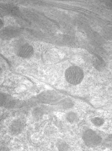
| Description | Zero degree tilt image of a 0.5 micron section of a mitochondrion and a mitochondrion-associated adherens complex in the Calyx of Held of the adult cat |
Embed
Embed URL
Embed Image
Full resolution data file
| File Size |
|
| File Format |
|
| Description | Tar.gz file (tilt_images.tar.gz) of the aligned tilt images of the the Mitochondrion-associated Adherens Complex (MAC) at the calyx of Held nerve terminal showing the location of mitochondria relative to the active sites and adherens junctions. |
Animation
Embed
Embed URL
Embed Video
| Description | Animation of aligned tilt images of the Mitochondrion-associated Adherens Complex (MAC) at the calyx of Held nerve terminal showing the location of mitochondria relative to the active sites and adherens junctions. |
Segmentation
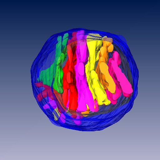
| Description | Segmentation of mitochondrion outer and inner membranes, cristae |
Embed
Embed URL
Embed Image
Full resolution data file
| File Size |
|
| File Format |
|
| Description | Tar.gz file (segment_data.tar.gz) of the segmentation of a mitochondrion from the Mitochondrion-associated Adherens Complex (MAC) at the calyx of Held nerve terminal showing the location of mitochondria relative to the active sites and adherens junctions. Shows outer membrane, inner membrane and cristae. |
Reconstruction
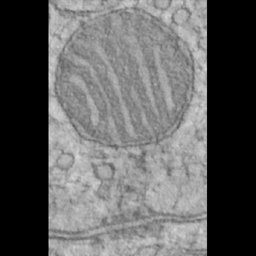
| Description | Conputed slice through a tomographic reconstruction of the mitochondrial adherens complex in the Calyx of Held of an adult cat. The specific mitochondrion is shown was segmented and can be found in the segmentation section of this record. |
Embed
Embed URL
Embed Image
Full resolution data file
| File Size |
|
| File Format |
|
| Description | Zip file containing the reconstruction file (king_filt.img/hdr) in Analyze 7.5 format (king_vol.zip) that was used to produce the segmentation for this reconstruction. |
Animation
Embed
Embed URL
Embed Video
| Description | Slices through the reconstructed volume |
Full metadata
- Collection
- Cite This Work
-
Perkins, Guy A.; Spirou, George (2017). Microscopy product ID: 3487. In Cell Centered Database. UC San Diego Library Digital Collections. https://doi.org/10.6075/J0DV1JPR
- Creation Date
- Project: 2003-06-01 to 2010-01-20.
- Date Issued
- 2017
- Research Team Head
- Researcher
- Technical Details
-
Male
Product type: SINGLE TILT. Microscopy type: IVEM. Instrument: JEOL 4000EX
- Funding
-
National Institutes of Health
- Series
- Scientific Name
- Anatomy
- Topic
Formats
View formats within this collection
- Language
- No linguistic content; Not applicable
- Related Resources
- Perkins GA, Tjong J, Brown JM, Poquiz PH, Scott RT, Kolson DR, Ellisman MH, Spirou GA (2010). The micro-architecture of mitochondria at active zones: electron tomography reveals novel anchoring scaffolds and cristae structured for high-rate metabolism. J Neurosci, 30(3):1015-1026. PubMed: https://www.ncbi.nlm.nih.gov/pmc/articles/PMC2829299/; Journal of Neuroscience. https://doi.org/10.1523/JNEUROSCI.1517-09.2010
- Microscopy product 3487 at the Cell Centered Database: https://doi.org/10.7295/W9CCDB3487
Primary associated publication
Other version
- License
-
Creative Commons Attribution 4.0 International Public License
- Rights Holder
- UC Regents
- Copyright
-
Under copyright (US)
Use: This work is available from the UC San Diego Library. This digital copy of the work is intended to support research, teaching, and private study.
Constraint(s) on Use: This work is protected by the U.S. Copyright Law (Title 17, U.S.C.). Use of this work beyond that allowed by "fair use" or any license applied to this work requires written permission of the copyright holder(s). Responsibility for obtaining permissions and any use and distribution of this work rests exclusively with the user and not the UC San Diego Library. Inquiries can be made to the UC San Diego Library program having custody of the work.
- Digital Object Made Available By
-
Research Data Curation Program, UC San Diego, La Jolla, 92093-0175 (https://lib.ucsd.edu/rdcp)
- Last Modified
2023-05-22