Microscopy product ID: 61937
2D Image
| Description | Zero degree tilt image of a presynaptic bouton from a cultured neuron expressing alpha synuclein-MiniSOG processed for photo-oxidation. Extensive stained membranous elements are observed in the pre-synaptic terminal. Data shown in Fig. 4B of Boassa et al., 2013. Use the brightness and contrast controls to adjust image. |
Embed
Embed URL
Embed Image
Full resolution data file
| File Size |
|
| File Format |
|
| Description | 463 |
Segmentation
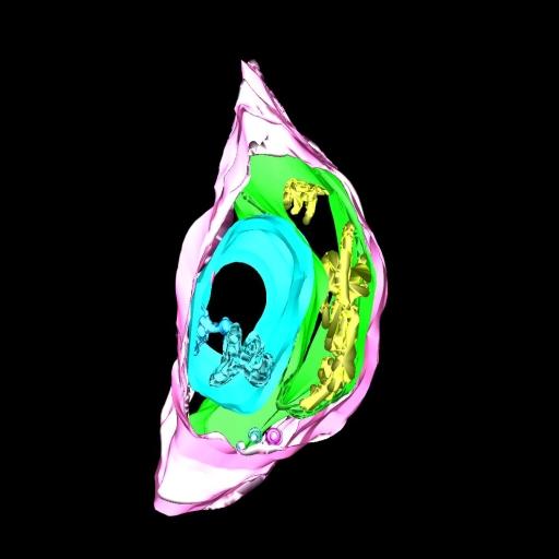
| Description | Manual segmentation of plasma membrane and labeled tubero-membranous system in a synaptic terminal |
Embed
Embed URL
Embed Image
Full resolution data file
| File Size |
|
| File Format |
|
| Description | Three-dimensional model from EM tomogram of same area as 4B shows plasma membrane (pink) and contiguous membranes in three different colors (blue, green, and yellow). |
Reconstruction
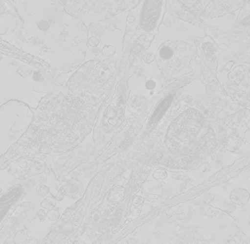
| Description | Computed slices through a double tilt electron tomographic reconstruction of synaptic terminals in rat cultured cortical neurons expressing alpha synuclein-MiniSOG and processed for photo-oxidation. Extensive stained membranous elements are observed in the pre-synaptic terminal Data corresponds to Fig. 4 in Boassa et al. 2013. |
Embed
Embed URL
Embed Image
Full resolution data file
| File Size |
|
| File Format |
|
| Description | Computed slices through a double tilt electron tomographic reconstruction of synaptic terminals in rat cultured cortical neurons expressing alpha synuclein-MiniSOG and processed for photo-oxidation. Extensive stained membranous elements are observed in the pre-synaptic terminal Data corresponds to Fig. 4 (Tomogram of cultured neurons expressing AS-MiniSOG processed for photooxidation. Extensive stained membranous elements are observed in the presynaptic terminal.)in Boassa et al. 2013. |
Full metadata
- Collection
- Cite This Work
-
Boassa, Daniela; Berlanga, Monica L.; Yang, Mary Ann; Terada, Masako; Hu, Junru; Bushong, Eric A.; Hwang, Minju; Masliah, Eliezer; George, Julia M.; Ellisman, Mark H. (2017). Microscopy product ID: 61937. In Cell Centered Database. UC San Diego Library Digital Collections. https://doi.org/10.6075/J0M908GQ
- Creation Date
- Experiment: 2011-03-08.
- Date Issued
- 2017
- Research Team Head
- Researchers
- Technical Details
-
Product type: DOUBLE TILT. Microscopy type: IVEM. Instrument: JEOL 4000EX
Strain: Sprague Dawley
- Series
- Scientific Name
- Anatomy
- Topic
Formats
View formats within this collection
- Language
- No linguistic content; Not applicable
- Related Resources
- Boassa D, Berlanga ML, Yang MA, Terada M, Hu J, Bushong EA, Hwang M, Masliah E, George JM, Ellisman MH (2013). Mapping the subcellular distribution of alpha-synuclein in neurons using genetically encoded probes for correlated light and electron microscopy: Implications for Parkinson's disease pathogenesis. J Neurosci, 33(6):2605-2615. PubMed: https://www.ncbi.nlm.nih.gov/pmc/articles/PMC3711410/; Journal of Neuroscience. https://doi.org/10.1523/JNEUROSCI.2898-12.2013
- Microscopy product 61937 at the Cell Centered Database: https://doi.org/10.7295/W9CCDB61937
Primary associated publication
Other version
- License
-
Creative Commons Attribution 4.0 International Public License
- Rights Holder
- UC Regents
- Copyright
-
Under copyright (US)
Use: This work is available from the UC San Diego Library. This digital copy of the work is intended to support research, teaching, and private study.
Constraint(s) on Use: This work is protected by the U.S. Copyright Law (Title 17, U.S.C.). Use of this work beyond that allowed by "fair use" or any license applied to this work requires written permission of the copyright holder(s). Responsibility for obtaining permissions and any use and distribution of this work rests exclusively with the user and not the UC San Diego Library. Inquiries can be made to the UC San Diego Library program having custody of the work.
- Digital Object Made Available By
-
Research Data Curation Program, UC San Diego, La Jolla, 92093-0175 (https://lib.ucsd.edu/rdcp)
- Last Modified
2023-05-22