Microscopy product ID: 3696
2D Image
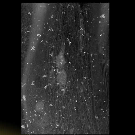
| Description | Single image taken on an intermediate voltage electron microscope at zero degrees tilt from a single axis tilt series through the Node of Ranvier from rat peripheral nerve prepared by chemical fixation followed by high pressure freezing and free substitution. Image is shown in negative contrast. Bright spots are colloidal gold particles applied to the section surface to serve as fiducial markers for subsequent alignment. |
Embed
Embed URL
Embed Image
Full resolution data file
| File Size |
|
| File Format |
|
| Description | Original tilt images in suprim format (*.f). Also contains fid, preali, rawtlt, st, suprim files needed for alignment. According to the scanning dimensions given in the paper, the images (hpf*.f) submitted represent cropped and aligned images used for the reconstruction. The raw tilt images are contained in the .st file as a multifile image stack. |
Segmentation
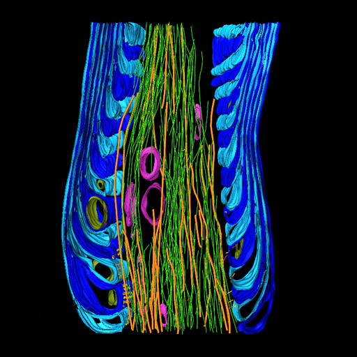
| Description | Manual segmentation of paranodal loops, microtubules, neurofilaments, sepjate junctions and cross bridges using Xvoxtrace v 2.9. Different parts of the myelin sheath were segmented as different objects. Objects were surfaced using both Synu and Amira |
Embed
Embed URL
Embed Image
Full resolution data file
| File Size |
|
| File Format |
|
| Description | Tar archive containing the trace file hpfnode_backup.trace and the segmented objects in synu (*.synu) and Amira (*.inventor) format. |
Reconstruction
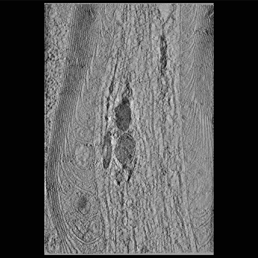
| Description | Single computed slice through a tomographic reconstruction of a Node of Ranvier from rat peripheral nerve, prepared by a combination of chemical fixation, high pressure freezing and freeze substitution |
Embed
Embed URL
Embed Image
Full resolution data file
| File Size |
|
| File Format |
|
| Description | Zip file contains the volume (yinghpf_good.img/hdr) in Analyze 7.5 format. |
Animation
Embed
Embed URL
Embed Video
| Description | Annotated movie through the computed slices of a tomographic reconstruction of a Node of Ranvier from rat peripheral nerve, along with surface renderings of structures segmented from the reconstruction. See the Segmentation section for a details about the structures and the segmentation process. |
Full metadata
- Collection
- Cite This Work
-
Ellisman, Mark H.; Sosinsky, Gina; Perkins, Guy A.; Ghassemzadeh, Sassan; Perez, Alex; Jones, Ying (2017). Microscopy product ID: 3696. In Cell Centered Database. UC San Diego Library Digital Collections. https://doi.org/10.6075/J0RX9BVB
- Date Issued
- 2017
- Research Team Heads
- Researchers
- Technical Details
-
Male. Strain: Sprague Dawley
Product type: SINGLE TILT. Microscopy type: IVEM. Instrument: JEOL 4000
- Funding
-
National Institutes of Health
- Series
- Scientific Name
- Anatomy
- Topic
Formats
View formats within this collection
- Language
- No linguistic content; Not applicable
- Related Resources
- Perkins GA, Sosinsky GE, Ghassemzadeh S, Perez A, Jones Y, Ellisman MH (2008). Electron tomographic analysis of cytoskeletal cross-bridges in the paranodal region of the node of Ranvier in peripheral nerves. J Struct Biol, 161(3):469-480. PubMed: https://www.ncbi.nlm.nih.gov/pmc/articles/PMC2346545/; Journal of Structural Biology. https://doi.org/10.1016/j.jsb.2007.10.005
- Microscopy product 3696 at the Cell Centered Database: https://doi.org/10.7295/W9CCDB3696
Primary associated publication
Other version
- License
-
Creative Commons Attribution 4.0 International Public License
- Rights Holder
- UC Regents
- Copyright
-
Under copyright (US)
Use: This work is available from the UC San Diego Library. This digital copy of the work is intended to support research, teaching, and private study.
Constraint(s) on Use: This work is protected by the U.S. Copyright Law (Title 17, U.S.C.). Use of this work beyond that allowed by "fair use" or any license applied to this work requires written permission of the copyright holder(s). Responsibility for obtaining permissions and any use and distribution of this work rests exclusively with the user and not the UC San Diego Library. Inquiries can be made to the UC San Diego Library program having custody of the work.
- Digital Object Made Available By
-
Research Data Curation Program, UC San Diego, La Jolla, 92093-0175 (https://lib.ucsd.edu/rdcp)
- Last Modified
2023-05-22