Microscopy product ID: 6624
2D Image
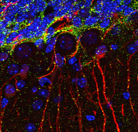
| Description | A representative tile from a large mosaic: this thumbnail image is a projection of 4 Z-slices from 3 fluorescent channels acquired from brain tissue using a 2-photon confocal microscope. Red is Rhodamine RedX labeling of Chicken anti-GFAP. Green is Alexafluor488 labeling of Connexin43. Blue is DAPI staining of all nuclei. |
Embed
Embed URL
Embed Image
Full resolution data file
| File Size |
|
| File Format |
|
| Description | This raw data file contains 7,104 3-color images (37x48x4) that make up the 1,776 tiles of this brain montage. A 50 micron thick sagittal section of the left hemisphere of the mouse brain was labeled with an antibody against Connexin43 protein and an antibody against GFAP protein. The nuclei of the brain tissue were labelled with DAPI staining. The area of the entire cerebellum was imaged using a 2-photon confocal microscope. |
Reconstruction
| Description | Large scale mosaic of mouse cerebellum reconstructed through automated acquisition and stitching of over 1700 individual tiles, triple labeled for GFAP (Red), Connexin 43 (Green) and DAPI (Blue), and imaged using multiphoton microscopy. The contrast and brightness was adjusted on the thumbnail image for display purposes and is different on the downloadable and Zoomify data. |
Embed
Embed URL
Embed Image
Full resolution data file
| File Size |
|
| File Format |
|
| Description | 1,776 tiled images were stitched together to reconstruct this single, high-resolution mosaic image of the cerebellum within a sagittal section of the left hemisphere of the mouse brain. Red is Rhodamine RedX labeling of Chicken anti-GFAP. Green is Alexafluor488 labeling of Rabbit anti-Connexin43. Blue is DAPI staining of all nuclei. |
Full metadata
- Collection
- Cite This Work
-
Cone, Angela; Sosinsky, Gina; Martone, Maryann (2017). Microscopy product ID: 6624. In Cell Centered Database. UC San Diego Library Digital Collections. https://doi.org/10.6075/J01C1WQ3
- Creation Date
- Microscopy product: 2008-12-30. Experiment: 2008-12-22. Project: 2008-11-06.
- Date Issued
- 2017
- Research Team Head
- Researchers
- Technical Details
-
Product type: MOSAIC. Microscopy type: multiphoton. Instrument: RTS2000
- Series
- Scientific Name
- Anatomy
- Topic
Formats
View formats within this collection
- Language
- No linguistic content; Not applicable
- Related Resource
- Microscopy product 6624 at the Cell Centered Database: https://doi.org/10.7295/W9CCDB6624
Other version
- License
-
Creative Commons Attribution 4.0 International Public License
- Rights Holder
- UC Regents
- Copyright
-
Under copyright (US)
Use: This work is available from the UC San Diego Library. This digital copy of the work is intended to support research, teaching, and private study.
Constraint(s) on Use: This work is protected by the U.S. Copyright Law (Title 17, U.S.C.). Use of this work beyond that allowed by "fair use" or any license applied to this work requires written permission of the copyright holder(s). Responsibility for obtaining permissions and any use and distribution of this work rests exclusively with the user and not the UC San Diego Library. Inquiries can be made to the UC San Diego Library program having custody of the work.
- Digital Object Made Available By
-
Research Data Curation Program, UC San Diego, La Jolla, 92093-0175 (https://lib.ucsd.edu/rdcp)
- Last Modified
2023-05-22