Microscopy product ID: 49
2D Image
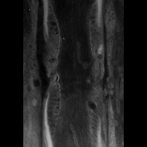
| Description | Zero tilt image through a 0.500 um section of the Node of Ranvier |
Embed
Embed URL
Embed Image
Segmentation
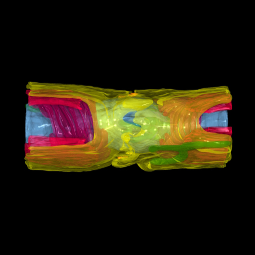
| Description | Manual segmentation of axonal and glial components of the Node of Ranvier using Xvoxtrace v 2.9. Different parts of the myelin sheath were segmented as different objects. Objects were surfaced using both Synu and Amira |
Embed
Embed URL
Embed Image
Full resolution data file
| File Size |
|
| File Format |
|
| Description | Zip archive containing the trace file node_marge2.trace and the segmented objects in synu (*.synu) and Amira (*.iv) format. Also included are several renderings of the surface model. These segmentations were produced from the mirrored volume file to mimic a complete node. That is, the original reconstruction of a sagittal section through a node was mirrored to produce the appearance of a single complete node. |
Reconstruction
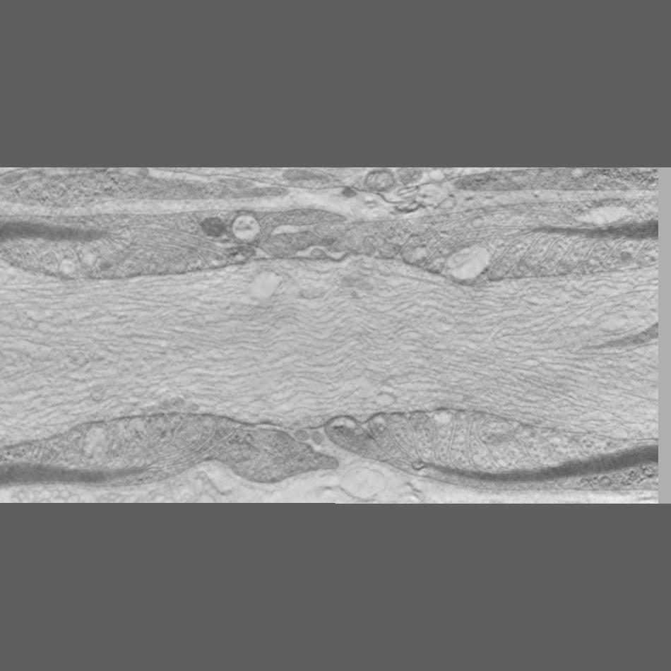
| Description | Single slice through a single tilt tomogram of the Node of Ranvier from mouse sciatic nerve prepared by high pressure freezing and freeze substitution of aldehyde fixed material |
Embed
Embed URL
Embed Image
Full resolution data file
| File Size |
|
| File Format |
|
| Description | Serial tomographic reconstruction from 3 serial volumes. The resulting volume was mirrored and merged with the original to from a relatively complete node of Ranvier. The downloadable zip file contains the merged volume in Analyze 7.5 format |
Animation
Embed
Embed URL
Embed Video
| Description | Annotated movie traversing the slices of the serial tomogram and identifying the segmented objects in the surface rendering |
Full metadata
- Collection
- Cite This Work
-
Ellisman, Mark H.; Sosinsky, Gina (2017). Microscopy product ID: 49. In Cell Centered Database. UC San Diego Library Digital Collections. https://doi.org/10.6075/J03F4PFG
- Creation Date
- Microscopy product: 2000-09-26. Experiment: 2000-09-26. Project: 1995-08-01 to 2006-06-30.
- Date Issued
- 2017
- Research Team Heads
- Technical Details
-
Male
Product type: SINGLE TILT. Microscopy type: IVEM. Instrument: JEOL 4000
- Funding
-
NIH NINDS
- Series
- Scientific Name
- Anatomy
- Topic
Formats
View formats within this collection
- Language
- No linguistic content; Not applicable
- Related Resources
- Sosinsky GE, Deerinck TJ, Greco R, Buitenhuys CH, Bartol TM, Ellisman MH (2005). Development of a model for microphysiological simulations: small nodes of ranvier from peripheral nerves of mice reconstructed by electron tomography. Neuroinformatics, 3(2):133-62. PubMed: https://www.ncbi.nlm.nih.gov/pubmed/15988042; Neuroinformatics. https://doi.org/10.1385/NI:3:2:133
- Microscopy product 49 at the Cell Centered Database: https://doi.org/10.7295/W9CCDB49
Primary associated publication
Other version
- License
-
Creative Commons Attribution 4.0 International Public License
- Rights Holder
- UC Regents
- Copyright
-
Under copyright (US)
Use: This work is available from the UC San Diego Library. This digital copy of the work is intended to support research, teaching, and private study.
Constraint(s) on Use: This work is protected by the U.S. Copyright Law (Title 17, U.S.C.). Use of this work beyond that allowed by "fair use" or any license applied to this work requires written permission of the copyright holder(s). Responsibility for obtaining permissions and any use and distribution of this work rests exclusively with the user and not the UC San Diego Library. Inquiries can be made to the UC San Diego Library program having custody of the work.
- Digital Object Made Available By
-
Research Data Curation Program, UC San Diego, La Jolla, 92093-0175 (https://lib.ucsd.edu/rdcp)
- Last Modified
2023-05-22