Microscopy product ID: 3742
2D Image
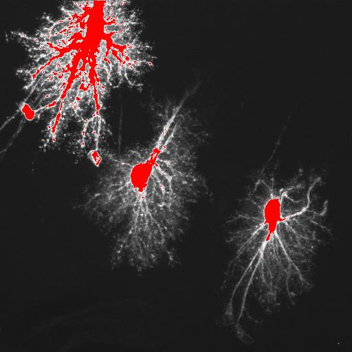
| Description | Projection through optical section series of filled astrocyte in the adult rat dentate gyrus. Saturated pixels are displayed in red. This section was also double labeled for EphA4 (see merged image under reconstruction). |
Embed
Embed URL
Embed Image
Full resolution data file
| File Size |
|
| File Format |
|
| Description | Zip file containing original optical section series through an intracellularly injected protoplasmic astrocyte (4s13as2_ly.pic) in dentate gyrus immunolabeled for EphA4 (4s13as2_eph.pic). Each label is in a separate file. The merged file is available for download under "Reconstruction." |
Animation
Embed
Embed URL
Embed Video
| Description | Animation through optical section series of a filled astrocyte in the molecular layer of the rat dentate gyrus imaged using confocal microscopy. Tissue was immunolabeled for EphA4. Animation for the merged data is available under "Reconstruction" |
Reconstruction
| Description | Projection through merged optical section series of a filled astrocyte (green) in the molecular layer of the dentate gyrus, stained for Epha4 (red), showing the relationship between astrocyte processes and laminar boundaries revealed by EphA4 staining. |
Embed
Embed URL
Embed Image
Full resolution data file
| File Size |
|
| File Format |
|
| Description | Zip file containing the merged channel file in tiff format. 4sl3as2_merger.tif.zip |
Animation
Embed
Embed URL
Embed Video
| Description | Animation through the optical sections of a confocal data set showing the relationship of a filled astrocyte (green) to laminar boundaries in the dentate gyrus revealed by EphA4 immunolabeling. The animation has been downsampled from the original data for ease of display. |
Full metadata
- Collection
- Cite This Work
-
Bushong, Eric A.; Martone, Maryann; Ellisman, Mark H. (2017). Microscopy product ID: 3742. In Cell Centered Database. UC San Diego Library Digital Collections. https://doi.org/10.6075/J0XG9QXV
- Creation Date
- Project: 2000-03-01 to 2003-07-23.
- Date Issued
- 2017
- Research Team Head
- Researchers
- Technical Details
-
Male. Strain: Sprague Dawley
Product type: OPTICAL SECTION. Microscopy type: laser scanning confocal. Instrument: BioRad 1024 MRC Confocal
- Funding
-
National Institutes of Health
- Series
- Scientific Name
- Anatomy
- Topic
Formats
View formats within this collection
- Language
- No linguistic content; Not applicable
- Related Resources
- Bushong EA, Martone ME, Ellisman MH (2003). Examination of the relationship between astrocyte morphology and laminar boundaries in the molecular layer of adult dentate gyrus. J Comp Neurol, 462(2):241-51. PubMed: https://www.ncbi.nlm.nih.gov/pubmed/12794746; Journal of Comparative Neuroscience. https://doi.org/10.1002/cne.10728
- Microscopy product 3742 at the Cell Centered Database: https://doi.org/10.7295/W9CCDB3742
Primary associated publication
Other version
- License
-
Creative Commons Attribution 4.0 International Public License
- Rights Holder
- UC Regents
- Copyright
-
Under copyright (US)
Use: This work is available from the UC San Diego Library. This digital copy of the work is intended to support research, teaching, and private study.
Constraint(s) on Use: This work is protected by the U.S. Copyright Law (Title 17, U.S.C.). Use of this work beyond that allowed by "fair use" or any license applied to this work requires written permission of the copyright holder(s). Responsibility for obtaining permissions and any use and distribution of this work rests exclusively with the user and not the UC San Diego Library. Inquiries can be made to the UC San Diego Library program having custody of the work.
- Digital Object Made Available By
-
Research Data Curation Program, UC San Diego, La Jolla, 92093-0175 (https://lib.ucsd.edu/rdcp)
- Last Modified
2023-05-22