Microscopy product ID: 7796
2D Image
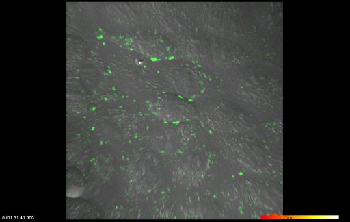
| Description | Merged fluorescent and DIC image of MDCK cells expressing Cx43-4C309/337 (green) |
Embed
Embed URL
Embed Image
Full resolution data file
| File Size |
|
| File Format |
|
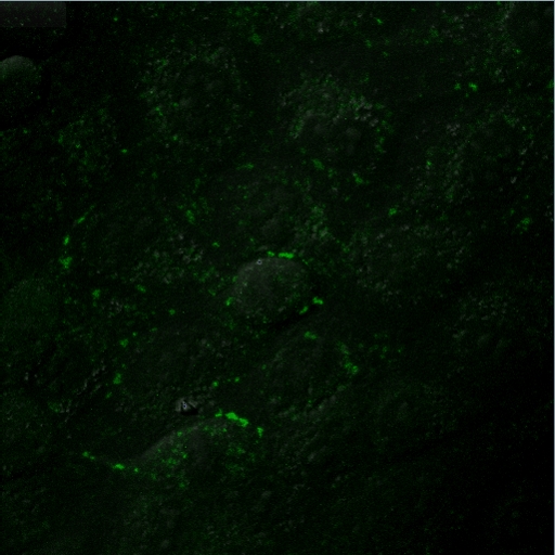
| Description | Optical section/time series in Olympus Fluoview .oif format. Each time point represents 11 z slices from the fluorescent channel and a DIC image. |
Animation
Embed
Embed URL
Embed Video
| Description | Time-lapse imaging of FlAsH-labeled MDCK cells expressing Cx43-4C309/337 (green) showing their rearrangement and fate during and after mitosis. FlAsH fluorescence excitation z series along with a transmitted DIC image were recorded every 6 min for a total duration of about 5 h. 3D volume reconstructions from confocal image stacks over time are displayed at a rate of two frames per minute. |
Full resolution data file
| File Size |
|
| File Format |
|
| Description | File containing the merged fluorescent-DIC imaging for each time point, where the fluorescent image is a maximum intensity projection of the z series collected at each time point. Each time point is represented as a "slice" in the volume in IMOD format. |
Animation
Embed
Embed URL
Embed Video
| Description | Annnotated time-lapse animation of FlAsH-labeled MDCK cells expressing Cx43-4C309/337 (green) showing their rearrangement and fate during and after mitosis. FlAsH fluorescence excitation z series along with a transmitted DIC image were recorded every 6 min for a total duration of about 5 h. 3D volume reconstructions from confocal image stacks over time are displayed at a rate of two frames per minute. Supplemental movie 2 from Boassa et al. (2010). |
Full metadata
- Collection
- Cite This Work
-
Boassa, Daniela; Sosinsky, Gina; Lampe, Paul; Solan, Joell (2017). Microscopy product ID: 7796. In Cell Centered Database. UC San Diego Library Digital Collections. https://doi.org/10.6075/J0J10300
- Date Issued
- 2017
- Research Team Head
- Researchers
- Technical Details
-
Product type: TIME SERIES. Microscopy type: LASER SCANNING CONFOCAL. Instrument: Olympus Fluoview 1000
- Funding
-
NIH GM072881 and GM065937 awarded to Gina Sosinky, NIH GM55632 awarded to Paul Lampe
- Series
- Scientific Name
- Anatomy
- Topic
Formats
View formats within this collection
- Language
- No linguistic content; Not applicable
- Related Resources
- Boassa D, Solan JL, Papas A, Thornton P, Lampe PD, Sosinsky GE (2010). Trafficking and recycling of the connexin43 gap junction protein during mitosis. Traffic, 11(11):1471-1486. PubMed: https://www.ncbi.nlm.nih.gov/pmc/articles/PMC3272544/; Traffic. https://doi.org/10.1111/j.1600-0854.2010.01109.x
- Microscopy product 7796 at the Cell Centered Database: https://doi.org/10.7295/W9CCDB7796
Primary associated publication
Other version
- License
-
Creative Commons Attribution 4.0 International Public License
- Rights Holder
- UC Regents
- Copyright
-
Under copyright (US)
Use: This work is available from the UC San Diego Library. This digital copy of the work is intended to support research, teaching, and private study.
Constraint(s) on Use: This work is protected by the U.S. Copyright Law (Title 17, U.S.C.). Use of this work beyond that allowed by "fair use" or any license applied to this work requires written permission of the copyright holder(s). Responsibility for obtaining permissions and any use and distribution of this work rests exclusively with the user and not the UC San Diego Library. Inquiries can be made to the UC San Diego Library program having custody of the work.
- Digital Object Made Available By
-
Research Data Curation Program, UC San Diego, La Jolla, 92093-0175 (https://lib.ucsd.edu/rdcp)
- Last Modified
2023-05-22